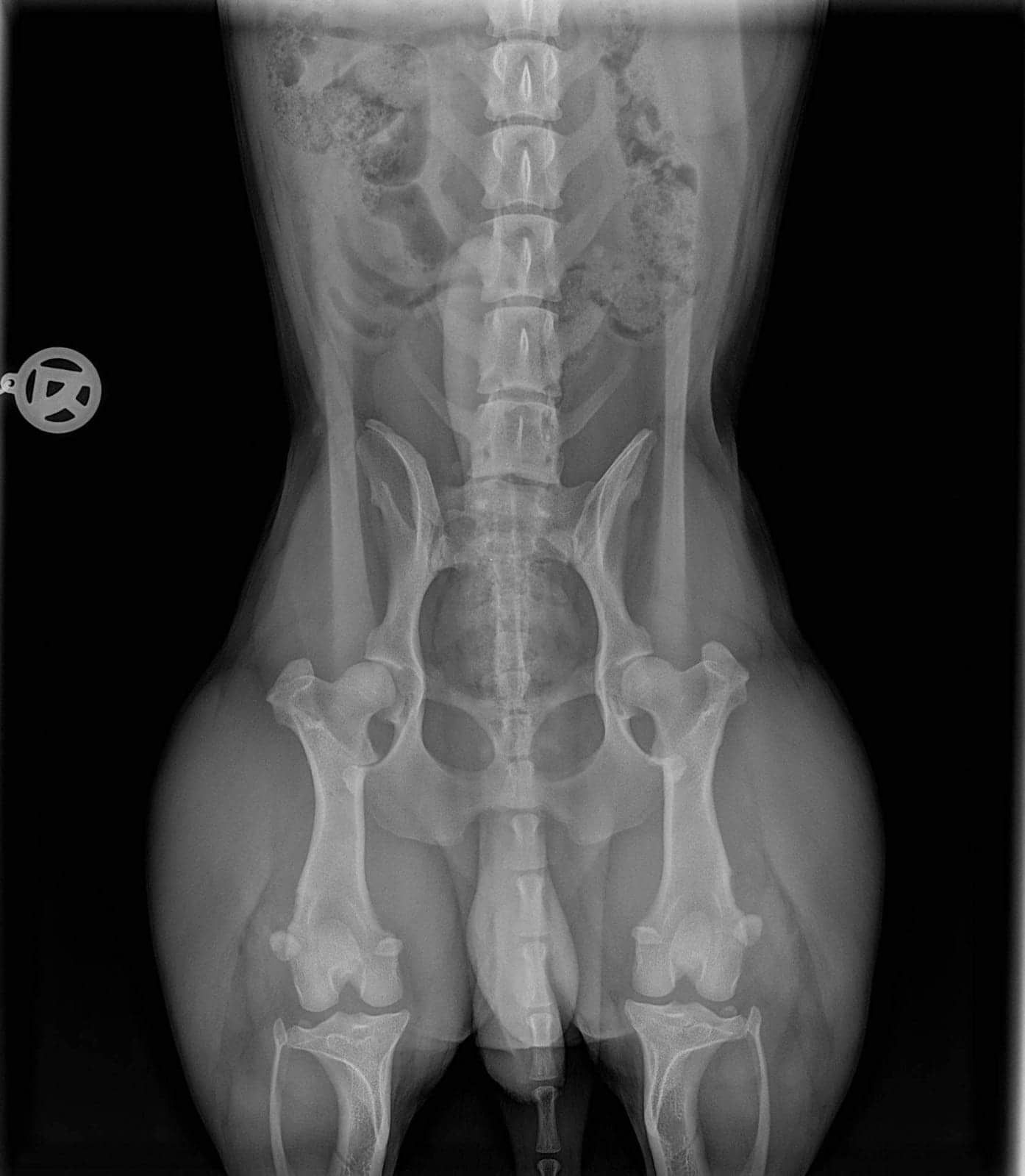
This document presents a detailed analysis of corgi hip x-rays, offering a systematic examination of the radiographic data collected from various subjects. The study is focused on the utilization of x-ray imaging to assess the anatomical structure and integrity of the hip joints in corgis, which is critical for the early identification and quantification of dysplastic changes.
The images have been evaluated using established radiographic protocols and standardized scoring systems, including the Orthopedic Foundation for Animals (OFA) criteria. These metrics provide a quantitative basis for assessing hip conformation and diagnosing potential anomalies. Through this analysis, the data aims to contribute to a more objective understanding of hip joint health and to support future research in veterinary orthopedics.
In the following sections, a curated list of hip x-rays is presented alongside pertinent observations and discussion of the imaging findings (added at a later point).
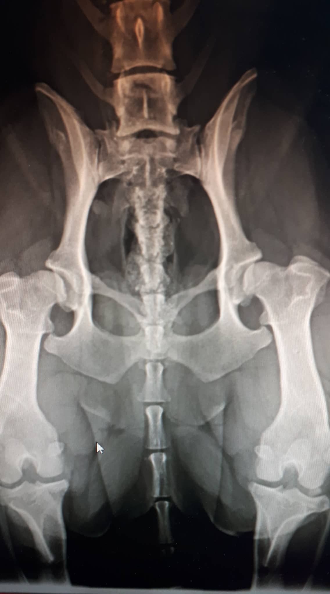
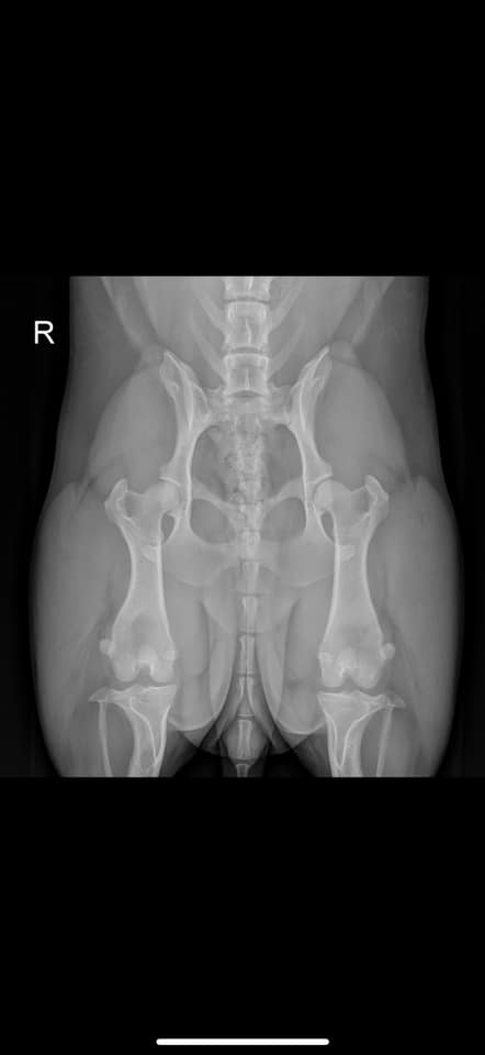
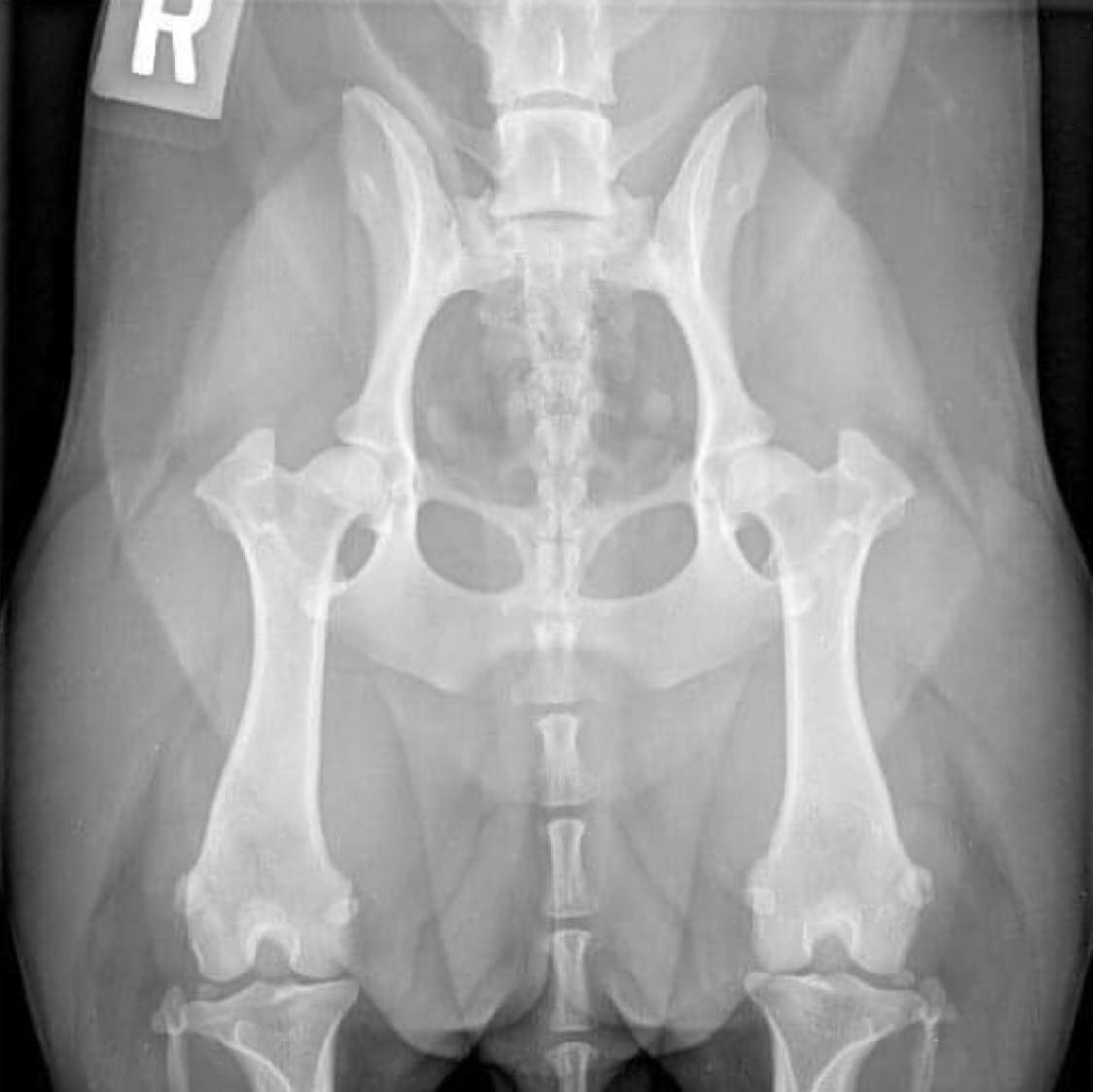
Excellent
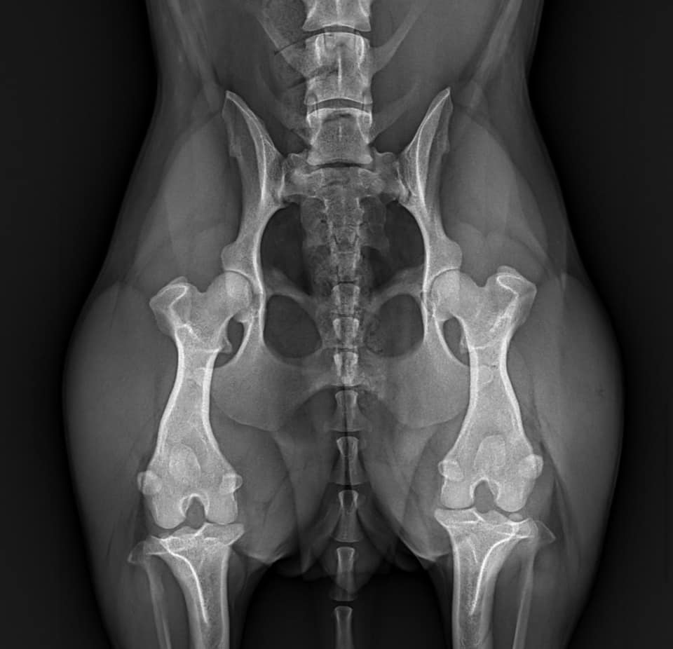

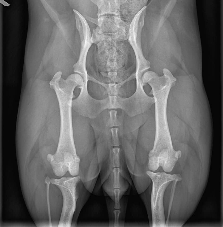
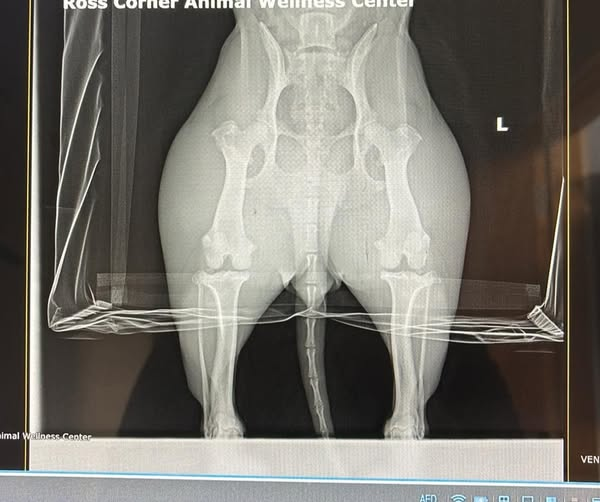
Good / Fair / Borderline (will be split up later)
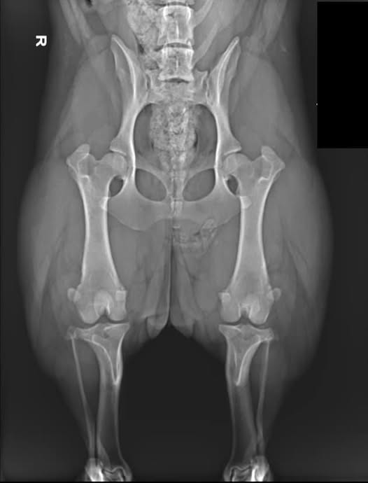
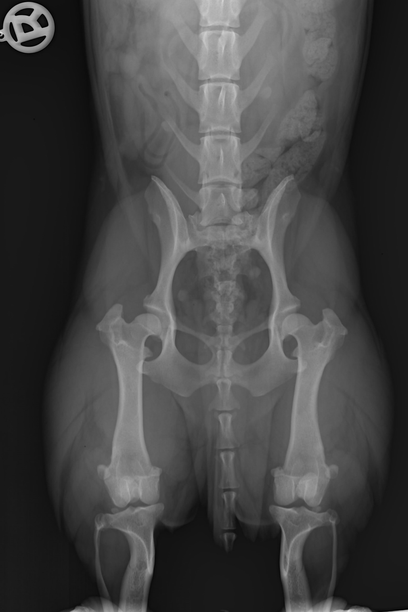
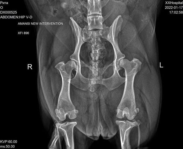
Borderline
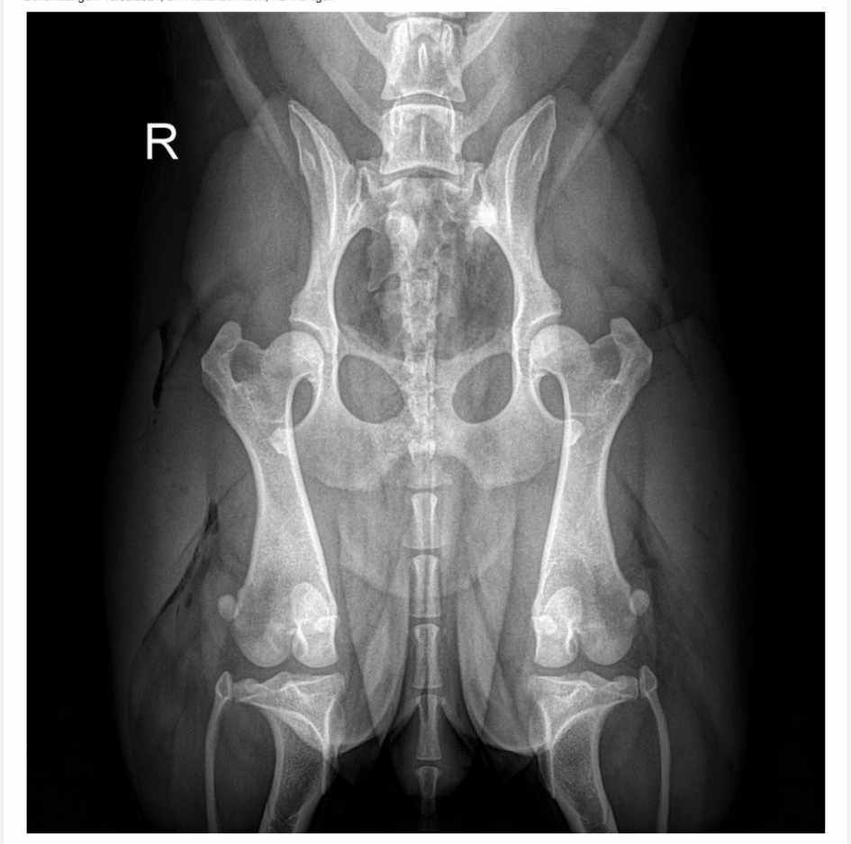
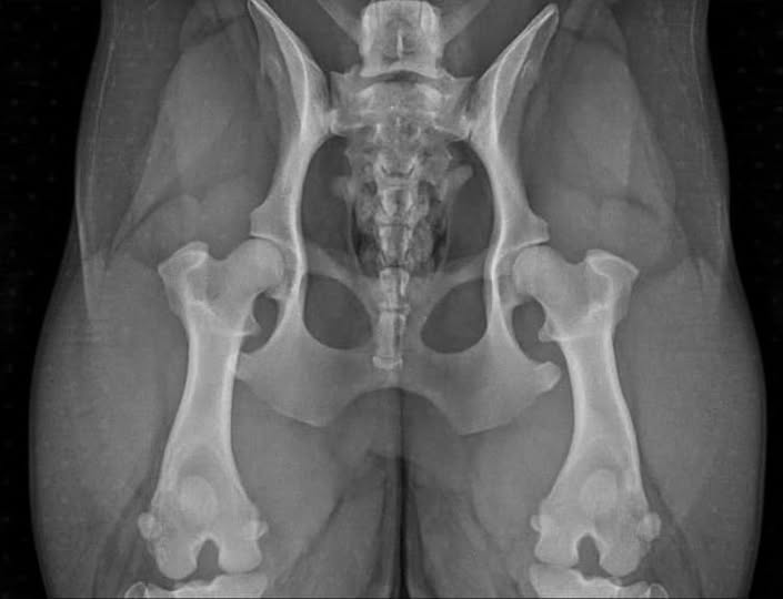
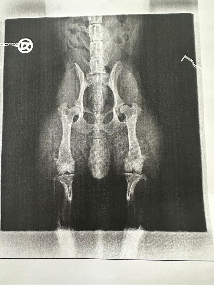
Mild / Moderate